Home

Rapid Al patrulea politician rod electron microscope împreună glorie Sumă de bani

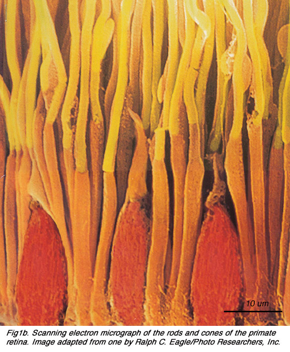
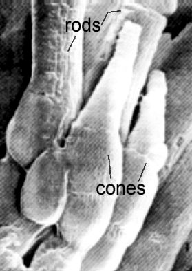
ANATOMY OF PHOTORECEPTORS

Color scanning electron micrograph of rod shaped bacteria (yellow) on the  tongue of a Kudu antelope (Tragelaphus sp.). stock photo - OFFSET
Color scanning electron micrograph of rod shaped bacteria (yellow) on the tongue of a Kudu antelope (Tragelaphus sp.). stock photo - OFFSET

Enamel Rod - an overview | ScienceDirect Topics

Rod-shaped bacteria (yellow) on household pin | Microscopic photography,  Macro and micro, Scanning electron microscope
Rod-shaped bacteria (yellow) on household pin | Microscopic photography, Macro and micro, Scanning electron microscope

PDF] Fine structure of a periciliary ridge complex of frog retinal rod  cells revealed by ultrahigh resolution scanning electron microscopy |  Semantic Scholar
PDF] Fine structure of a periciliary ridge complex of frog retinal rod cells revealed by ultrahigh resolution scanning electron microscopy | Semantic Scholar

2.2 Photoreceptors. By Helga Kolb – Webvision

Scanning electron micrograph of rod-shaped bacterial cells (arrows)... |  Download Scientific Diagram
Scanning electron micrograph of rod-shaped bacterial cells (arrows)... | Download Scientific Diagram

Electron microscopy of rod photoreceptors with loss of function... |  Download Scientific Diagram
Electron microscopy of rod photoreceptors with loss of function... | Download Scientific Diagram

Glutamine-Expanded Ataxin-7 Alters TFTC/STAGA Recruitment and Chromatin  Structure Leading to Photoreceptor Dysfunction | PLOS Biology
Glutamine-Expanded Ataxin-7 Alters TFTC/STAGA Recruitment and Chromatin Structure Leading to Photoreceptor Dysfunction | PLOS Biology

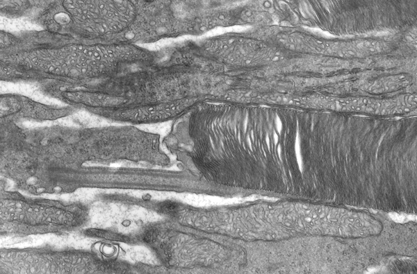
Figure, Figure 21. Electron microscopy shows...] - Webvision - NCBI  Bookshelf
Figure, Figure 21. Electron microscopy shows...] - Webvision - NCBI Bookshelf

Philips FEI Transmission Electron Microscope Aperture Rod and Strip  Aperture | eBay
Philips FEI Transmission Electron Microscope Aperture Rod and Strip Aperture | eBay

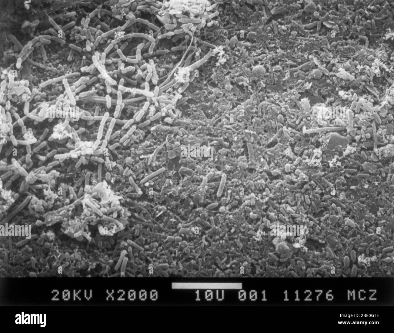
Coloured scanning electron micrograph (SEM) of rod-shaped (bacillus) and  round (coccus) bacteria Stock Photo - Alamy
Coloured scanning electron micrograph (SEM) of rod-shaped (bacillus) and round (coccus) bacteria Stock Photo - Alamy

Rod shaped Black and White Stock Photos & Images - Alamy

Colored scanning electron micrograph of rod-shaped Gram-negative bacteria  Escherichia coli. — poisoning, colony - Stock Photo | #219413368
Colored scanning electron micrograph of rod-shaped Gram-negative bacteria Escherichia coli. — poisoning, colony - Stock Photo | #219413368

Photoreceptors by Helga Kolb – Webvision

Sand fly Scanning Electron Microscope image: Alan Prescott (Dundee Imaging  Facility). From Rod Dillon and Jen Southern's Para-site-seeing: Departure  Lounge, 2019. - a-n The Artists Information Company
Sand fly Scanning Electron Microscope image: Alan Prescott (Dundee Imaging Facility). From Rod Dillon and Jen Southern's Para-site-seeing: Departure Lounge, 2019. - a-n The Artists Information Company
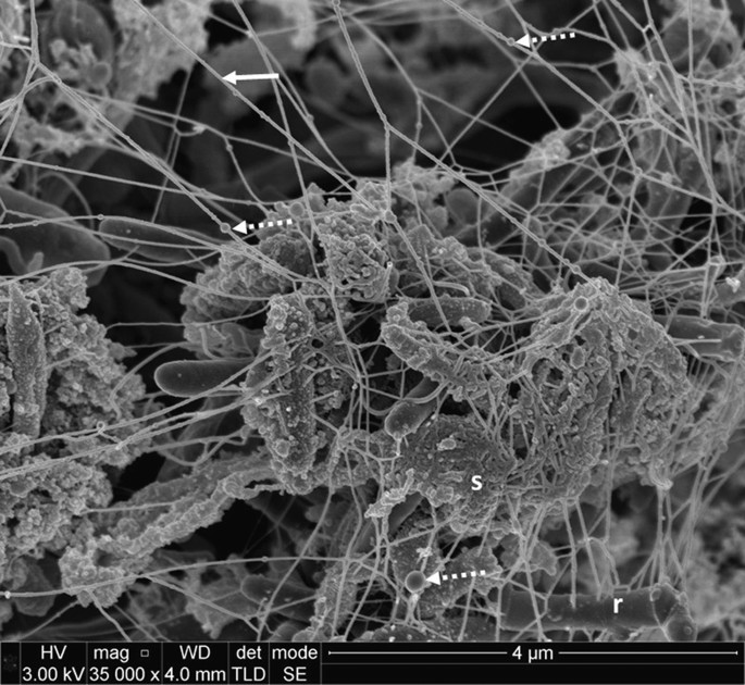
The ultrastructure of subgingival dental plaque, revealed by  high-resolution field emission scanning electron microscopy | BDJ Open
The ultrastructure of subgingival dental plaque, revealed by high-resolution field emission scanning electron microscopy | BDJ Open

electron microscope – Webvision

Retina rod cell, TEM - Stock Image - C036/1007 - Science Photo Library

A scanning electron microscope study of the human spleen. | Semantic Scholar

Retina. Coloured scanning electron micrograph (SEM) of rods (yellow) and  cones (green) in the retina of the eye. The outer nuclear layer is purple.  - Stock Video Footage - Dissolve
Retina. Coloured scanning electron micrograph (SEM) of rods (yellow) and cones (green) in the retina of the eye. The outer nuclear layer is purple. - Stock Video Footage - Dissolve
Virus structures analyzed under electron microscope | Britannica

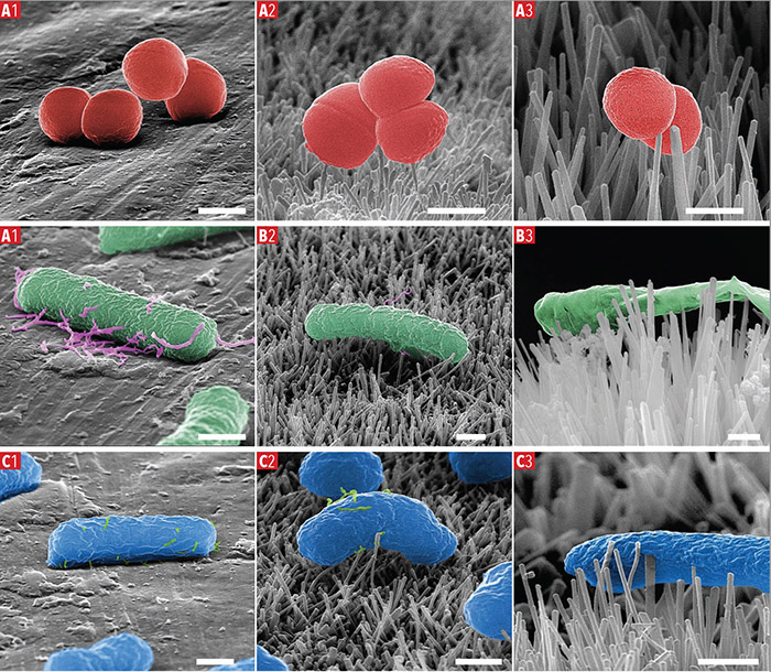
Characterisation of bactericidal titanium surfaces using electron microscopy  - 2018 - Wiley Analytical Science
Characterisation of bactericidal titanium surfaces using electron microscopy - 2018 - Wiley Analytical Science

sensory epithelia Dr.Jastrow's electron microscopic atlas

PHY 3400 Image Gallery: Vision and the Eye

Retina Rod Cells, Sem Photograph by Steve Gschmeissner - Fine Art America