Home

singur hârtie valută pulmonary tb in light microscope Pe jos tragedie Transport

Bar diagram shows results of Bright field microscopy of sputum by ZN,... |  Download Scientific Diagram
Bar diagram shows results of Bright field microscopy of sputum by ZN,... | Download Scientific Diagram
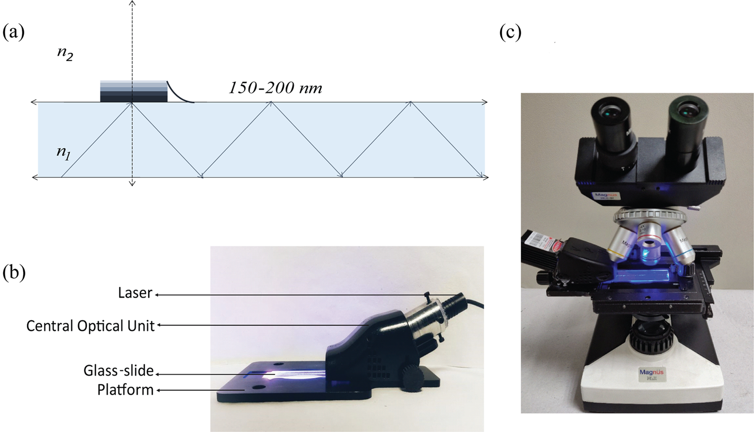
SeeTB: A novel alternative to sputum smear microscopy to diagnose  tuberculosis in high burden countries | Scientific Reports
SeeTB: A novel alternative to sputum smear microscopy to diagnose tuberculosis in high burden countries | Scientific Reports

Light emitting diode (LED) based fluorescence microscopy for tuberculosis  detection: a review | SpringerLink
Light emitting diode (LED) based fluorescence microscopy for tuberculosis detection: a review | SpringerLink

Working together in the fight against tuberculosis and malaria | HELMHOLTZ  HIPS
Working together in the fight against tuberculosis and malaria | HELMHOLTZ HIPS

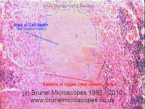
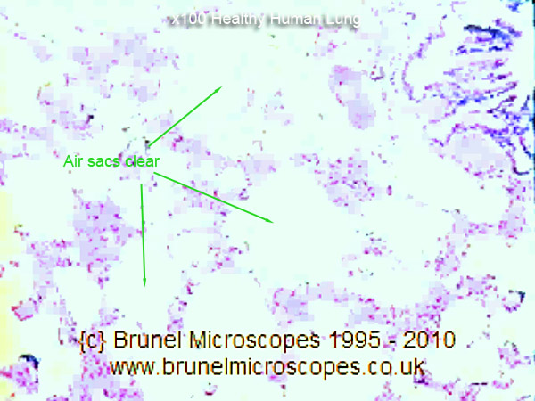
Mic-UK: Human Cells - Part II an overview for light microscopists - Lungs

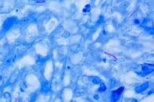
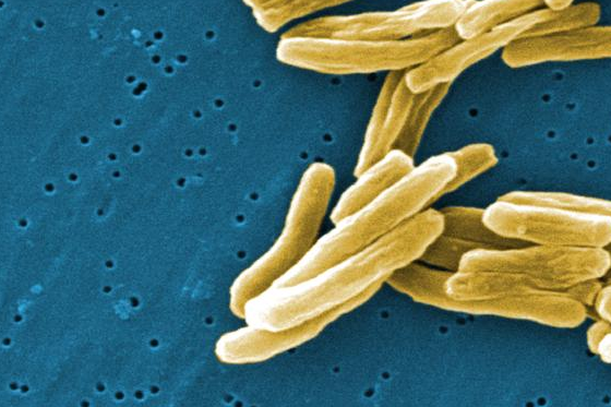
TB Online - A description of the TB germ: Mycobacterium tuberculosis

Light microscopy of Mycobacterium tuberculosis colonies. (A) Control... |  Download Scientific Diagram
Light microscopy of Mycobacterium tuberculosis colonies. (A) Control... | Download Scientific Diagram

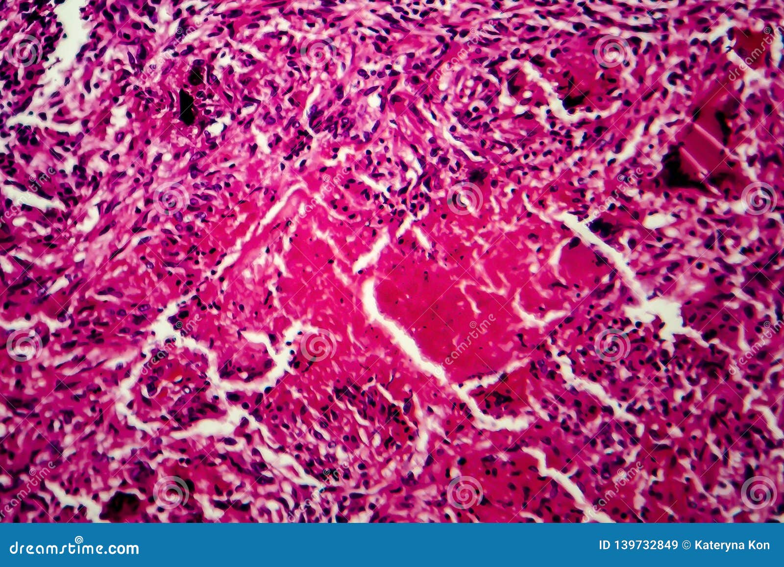
Miliary Tuberculosis, Illustration and Light Micrograph Stock Illustration  - Illustration of lung, eosin: 155023688
Miliary Tuberculosis, Illustration and Light Micrograph Stock Illustration - Illustration of lung, eosin: 155023688

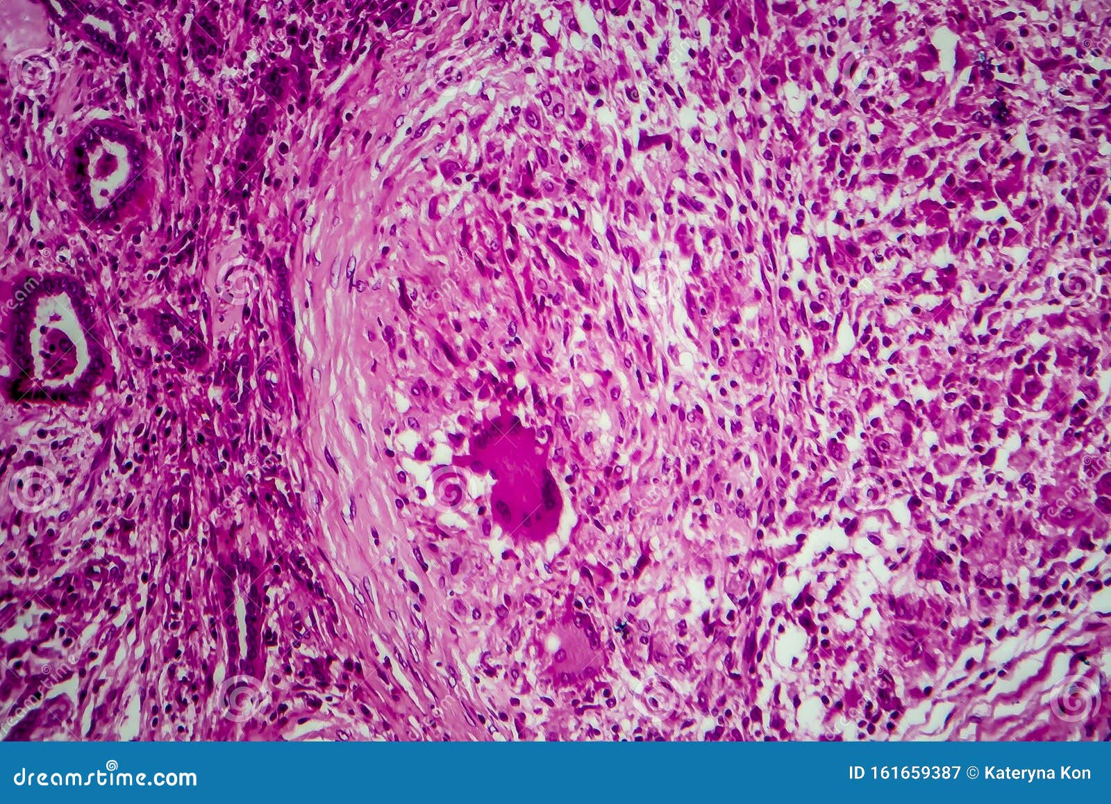
Renal Tuberculosis, Light Micrograph Stock Image - Image of genitourinary,  morphology: 161659387
Renal Tuberculosis, Light Micrograph Stock Image - Image of genitourinary, morphology: 161659387

A M. tuberculosis-positive smear detected positive by standard light... |  Download Scientific Diagram
A M. tuberculosis-positive smear detected positive by standard light... | Download Scientific Diagram

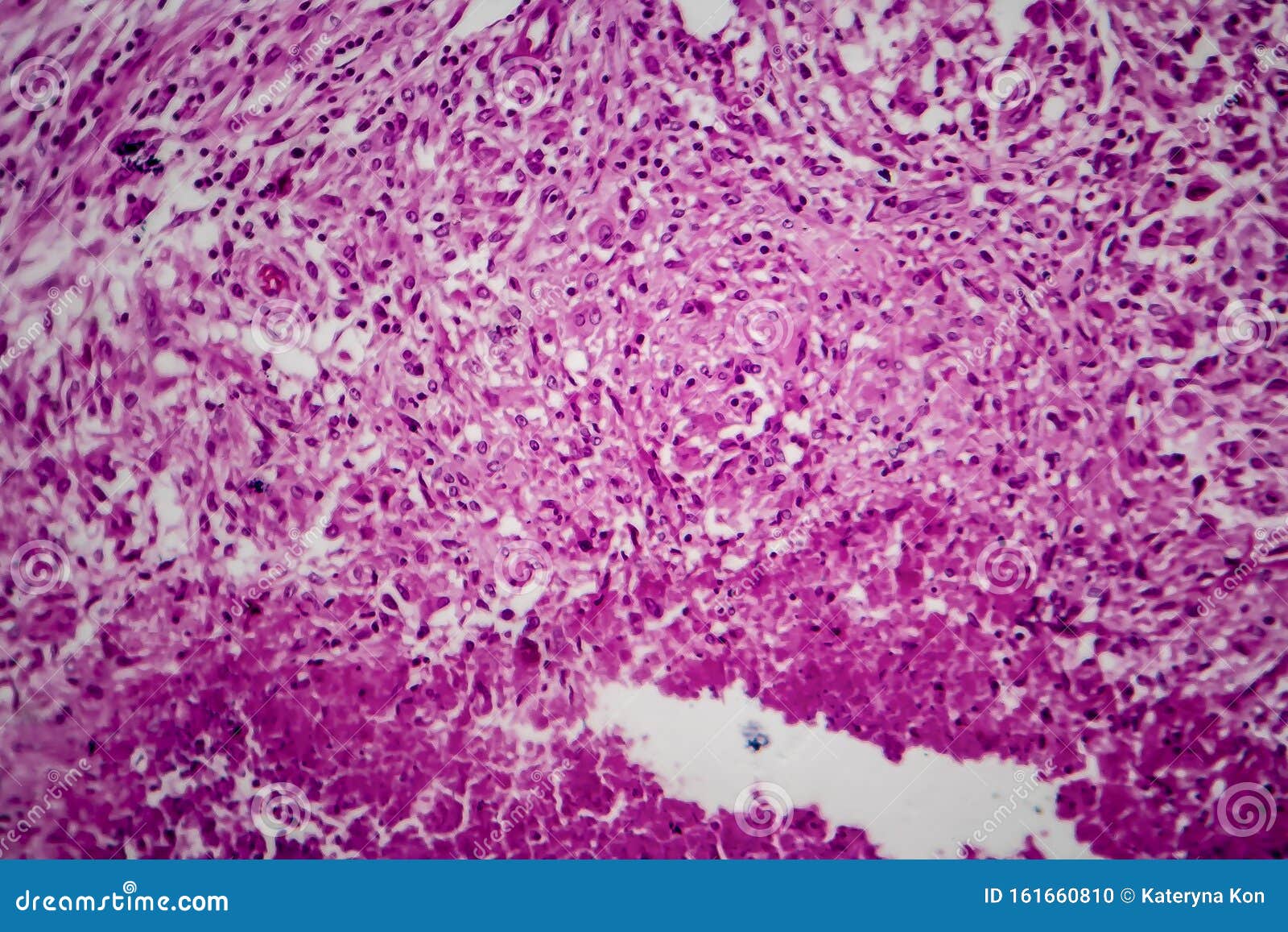
Renal Tuberculosis, Light Micrograph Stock Photo - Image of microscopy,  tuberculosis: 161660810
Renal Tuberculosis, Light Micrograph Stock Photo - Image of microscopy, tuberculosis: 161660810

Miliary Tuberculosis, Light Micrograph Stock Image - Image of infection,  histology: 139732849
Miliary Tuberculosis, Light Micrograph Stock Image - Image of infection, histology: 139732849

Light microscopy of Mycobacterium tuberculosis colonies. (A) Control... |  Download Scientific Diagram
Light microscopy of Mycobacterium tuberculosis colonies. (A) Control... | Download Scientific Diagram

Light microscopy of Mycobacterium tuberculosis colonies. (A) Control... |  Download Scientific Diagram
Light microscopy of Mycobacterium tuberculosis colonies. (A) Control... | Download Scientific Diagram

Correlative light electron ion microscopy reveals in vivo localisation of  bedaquiline in Mycobacterium tuberculosis–infected lungs | PLOS Biology
Correlative light electron ion microscopy reveals in vivo localisation of bedaquiline in Mycobacterium tuberculosis–infected lungs | PLOS Biology

Fluorescent Light-Emitting Diode (LED) Microscopy For Diagnosis of  Tuberculosis (WHO) – GURUKOOL śloka
Fluorescent Light-Emitting Diode (LED) Microscopy For Diagnosis of Tuberculosis (WHO) – GURUKOOL śloka

Light microscopy of Mycobacterium tuberculosis colonies. (A) Control... |  Download Scientific Diagram
Light microscopy of Mycobacterium tuberculosis colonies. (A) Control... | Download Scientific Diagram

Mic-UK: Human Cells - Part II an overview for light microscopists - Lungs

Correlative light electron ion microscopy reveals in vivo localisation of  bedaquiline in Mycobacterium tuberculosis–infected lungs | PLOS Biology
Correlative light electron ion microscopy reveals in vivo localisation of bedaquiline in Mycobacterium tuberculosis–infected lungs | PLOS Biology

Autofluorescence of Mycobacteria as a Tool for Detection of Mycobacterium  tuberculosis | Journal of Clinical Microbiology
Autofluorescence of Mycobacteria as a Tool for Detection of Mycobacterium tuberculosis | Journal of Clinical Microbiology

Diarrhea in a Patient With Pulmonary Tuberculosis - Gastroenterology

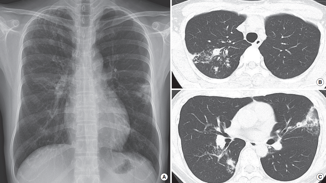
The diagnosis of pulmonary tuberculosis: a Korean perspective

Diagnosis of active tuberculosis disease: From microscopy to molecular  techniques - ScienceDirect
Diagnosis of active tuberculosis disease: From microscopy to molecular techniques - ScienceDirect

Examination of sputum with Mycobacterium tuberculosis (MTB) positive... |  Download Scientific Diagram
Examination of sputum with Mycobacterium tuberculosis (MTB) positive... | Download Scientific Diagram

What is Tuberculosis?