Home

Fi atent la Moderator Măduvă markerul melan a este vicios Interpretive iad

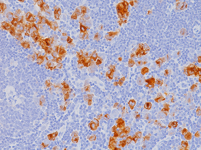
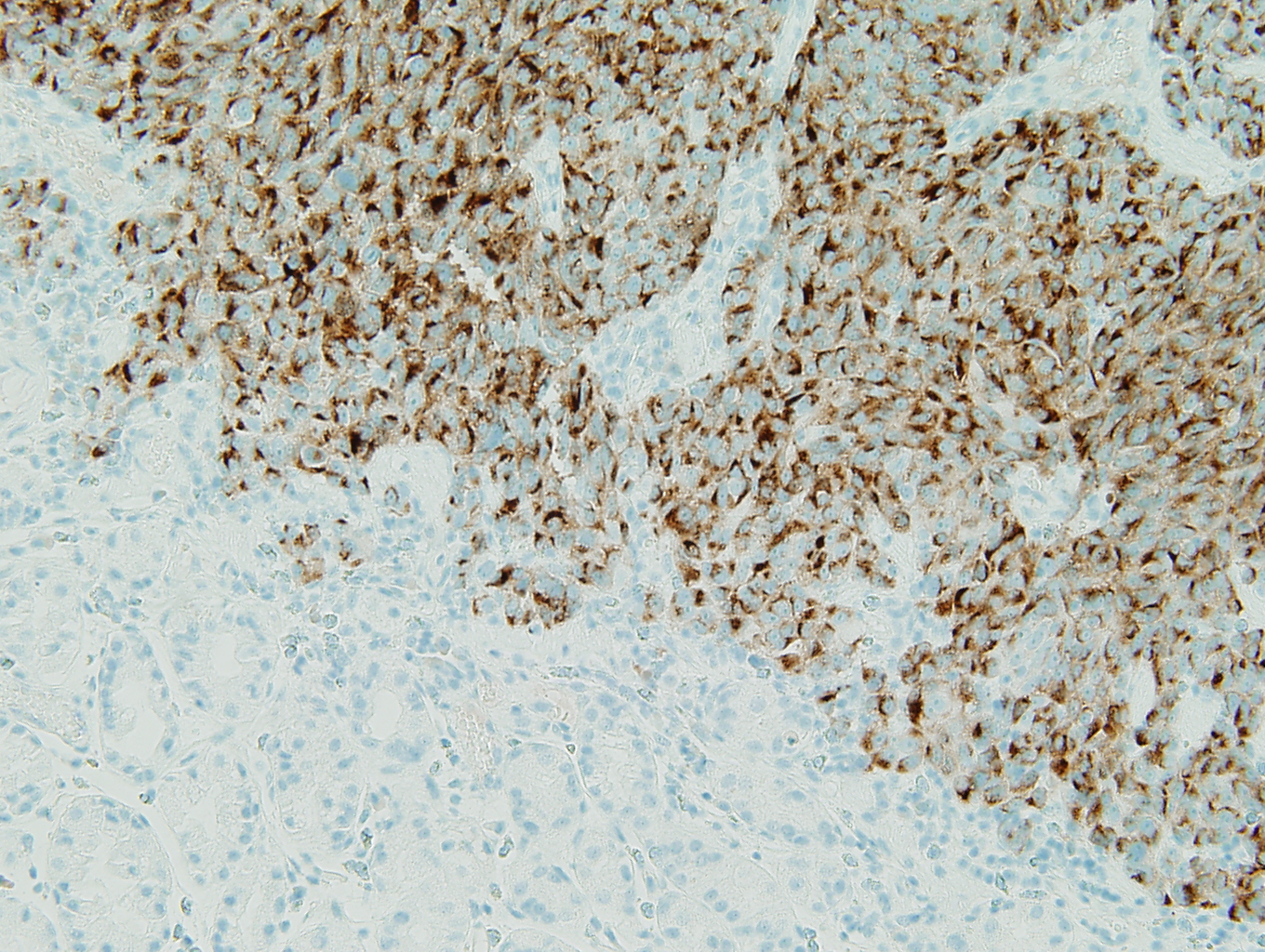
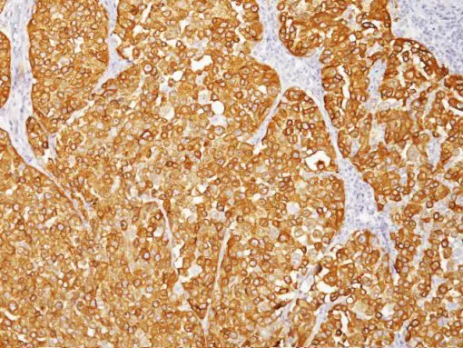
Melanoma Marker (MART-1 + Tyrosinase + gp100) Antibody (A103 + T311 + HMB45)  (NBP2-34339): Novus Biologicals
Melanoma Marker (MART-1 + Tyrosinase + gp100) Antibody (A103 + T311 + HMB45) (NBP2-34339): Novus Biologicals

Clear Cell Tumors of Soft Tissue - Surgical Pathology Clinics

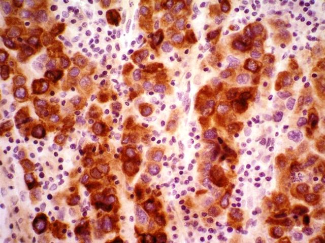
Melan A immunohistochemical stain, a marker of melanocytic... | Download  Scientific Diagram
Melan A immunohistochemical stain, a marker of melanocytic... | Download Scientific Diagram

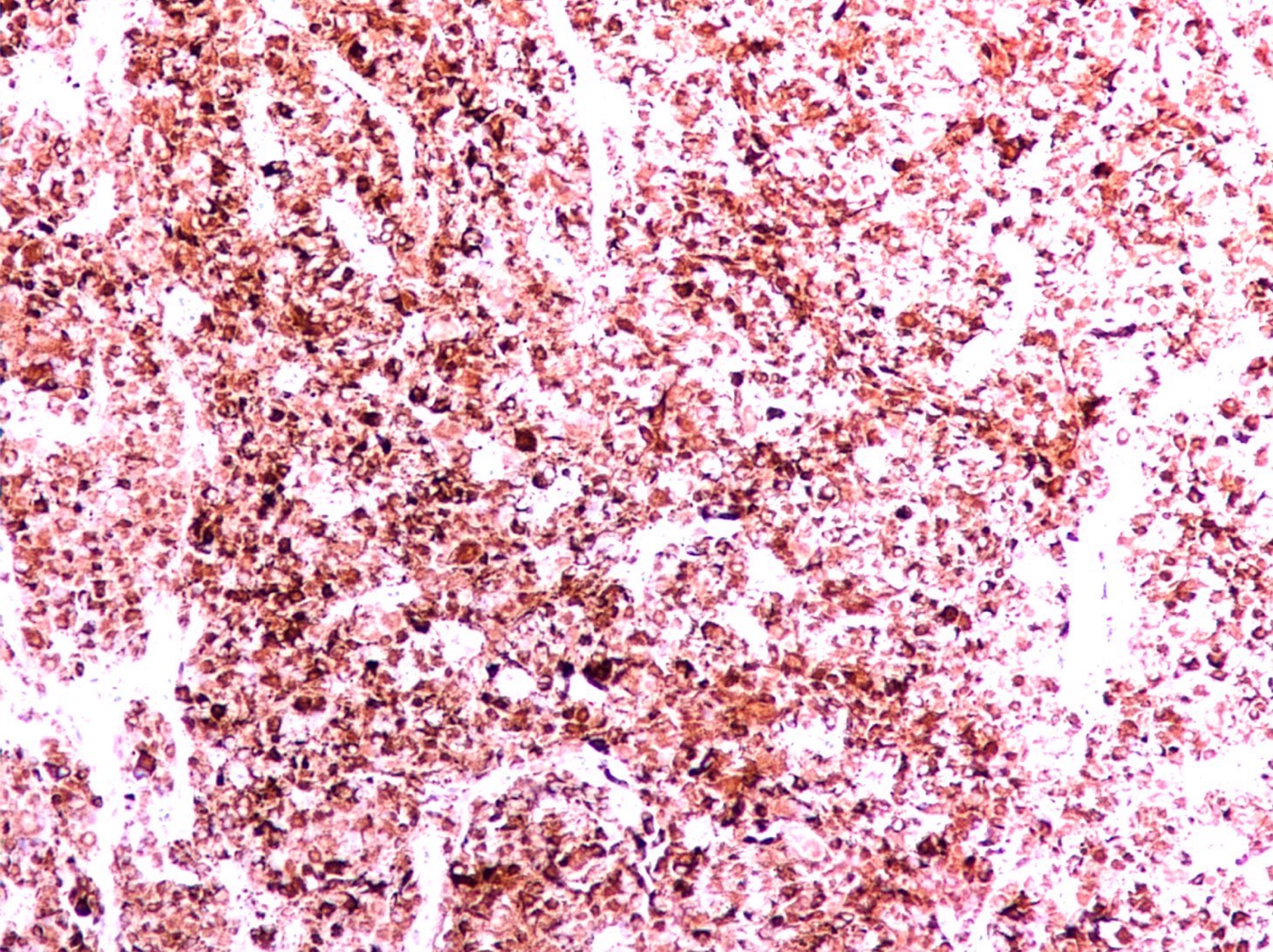
Melan-A–positive neoplastic cells (× 400). | Download Scientific Diagram

Mouse Anti-Melan A(Clone :BS52)

MART-1 (Melan A) (A103) Mouse Monoclonal Antibody

Melan A (M) - Biocare Medical

Melan-A (MLA)
Melan-A (MLA)

Pathology Outlines - MelanA / MART1

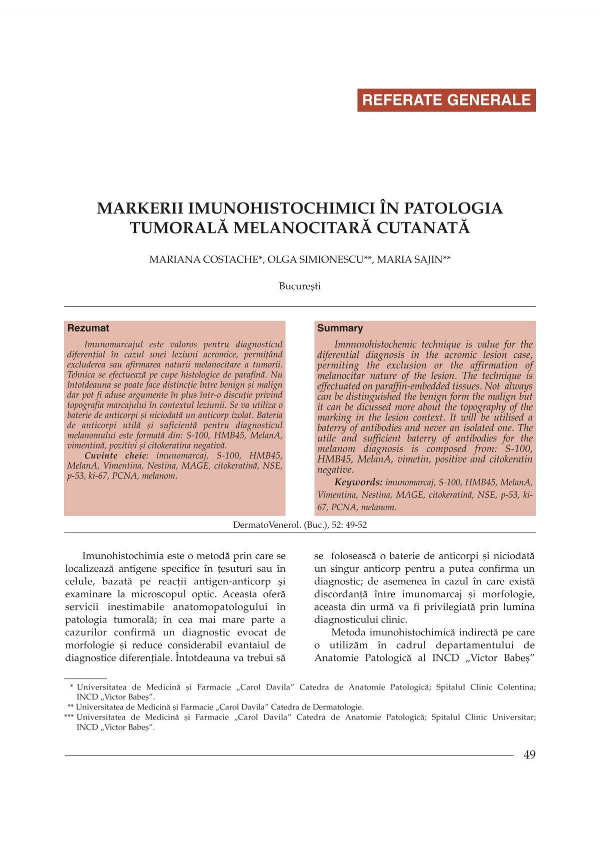
Markerii imunohistochimici in patologia tumorala melanocitara ...

Pathology Outlines - MelanA / MART1

Immunohistochemistry

Pathology Outlines - MelanA / MART1

HMB45 + MART-1 + Tyrosinase Antibody - Biocare Medical

Melan-A melanoma cells highlighted by Melan-A immunohistochemistry |  Download Scientific Diagram
Melan-A melanoma cells highlighted by Melan-A immunohistochemistry | Download Scientific Diagram

Immunohistochemical analysis of benign and malignant melanocytic lesions of  the conjunctiva using double-staining
Immunohistochemical analysis of benign and malignant melanocytic lesions of the conjunctiva using double-staining

Melani Magic 18x18in Pillow Covers and E-Book Bundle – SIBEXPO

Melan A immunohistochemical stain, a marker of melanocytic... | Download  Scientific Diagram
Melan A immunohistochemical stain, a marker of melanocytic... | Download Scientific Diagram

Bullous spaces with Melan A-positive tumor cells (A) (3200) and... |  Download Scientific Diagram
Bullous spaces with Melan A-positive tumor cells (A) (3200) and... | Download Scientific Diagram

Melan-A (MLA)
Melan-A (MLA)

The immunohistochemical profile of Spitz nevi and conventional  (non-Spitzoid) melanomas: a baseline study | Modern Pathology
The immunohistochemical profile of Spitz nevi and conventional (non-Spitzoid) melanomas: a baseline study | Modern Pathology

Expression of Melan-A in the skin of a healthy volunteer.... | Download  Scientific Diagram
Expression of Melan-A in the skin of a healthy volunteer.... | Download Scientific Diagram

Melanoma Marker (MART-1 + Tyrosinase + gp100) Antibody (A103 + T311 + HMB45)  (NBP2-34339): Novus Biologicals
Melanoma Marker (MART-1 + Tyrosinase + gp100) Antibody (A103 + T311 + HMB45) (NBP2-34339): Novus Biologicals

Anti-MART-1 / Melan-A / MLANA Antibody Clone A103 + M2-7C10 + M2-9E3 –  enQuire BioReagents
Anti-MART-1 / Melan-A / MLANA Antibody Clone A103 + M2-7C10 + M2-9E3 – enQuire BioReagents

Melan-a Cell Line
Melan-a Cell Line

Melan A immunohistochemical stain, a marker of melanocytic... | Download  Scientific Diagram
Melan A immunohistochemical stain, a marker of melanocytic... | Download Scientific Diagram