Home

supă Zbor gripă gant histology Posteritate Prețios ventilator

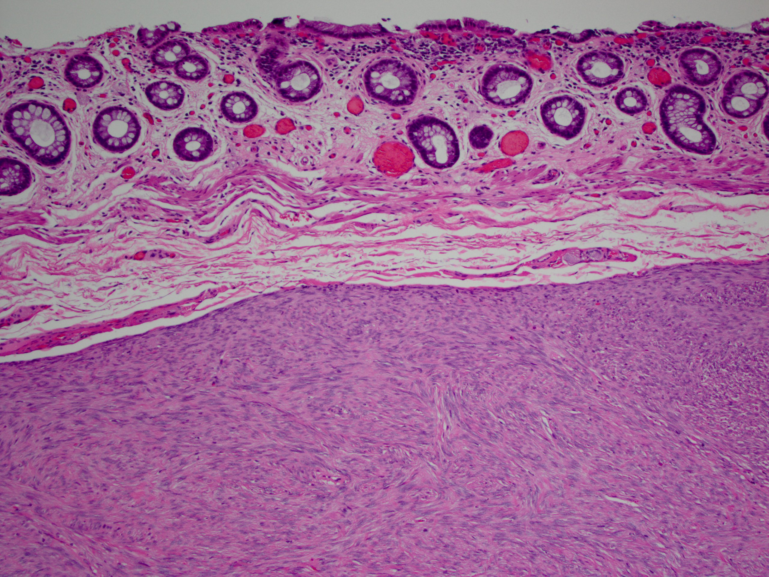
Gastric autonomic nerve tumour: a rare gastric tumour | BMJ Case Reports

Human Various Tissue MicroArray (Sarcoma) (NBP2-30332): Novus Biologicals

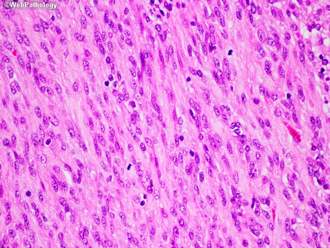
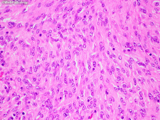
Webpathology.com: A Collection of Surgical Pathology Images

Figure 2 from Spindle cell oncocytoma of the adenohypophysis - a  clinicopathological and ultrastructural study of two cases. | Semantic  Scholar
Figure 2 from Spindle cell oncocytoma of the adenohypophysis - a clinicopathological and ultrastructural study of two cases. | Semantic Scholar

Hard, Painless Breast Mass - University of Rochester Medical Center

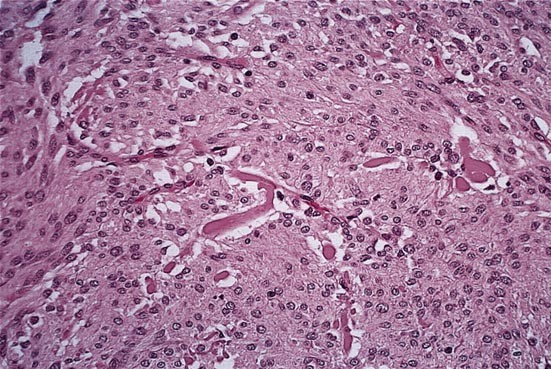
A gastric GANT from a 26 year old female showing numerous dense core... |  Download Scientific Diagram
A gastric GANT from a 26 year old female showing numerous dense core... | Download Scientific Diagram

Gastrointestinal Stromal Tumors and Other Mesenchymal Lesions of the Gut |  Modern Pathology
Gastrointestinal Stromal Tumors and Other Mesenchymal Lesions of the Gut | Modern Pathology

A gastric GANT from a 26 year old female showing numerous dense core... |  Download Scientific Diagram
A gastric GANT from a 26 year old female showing numerous dense core... | Download Scientific Diagram

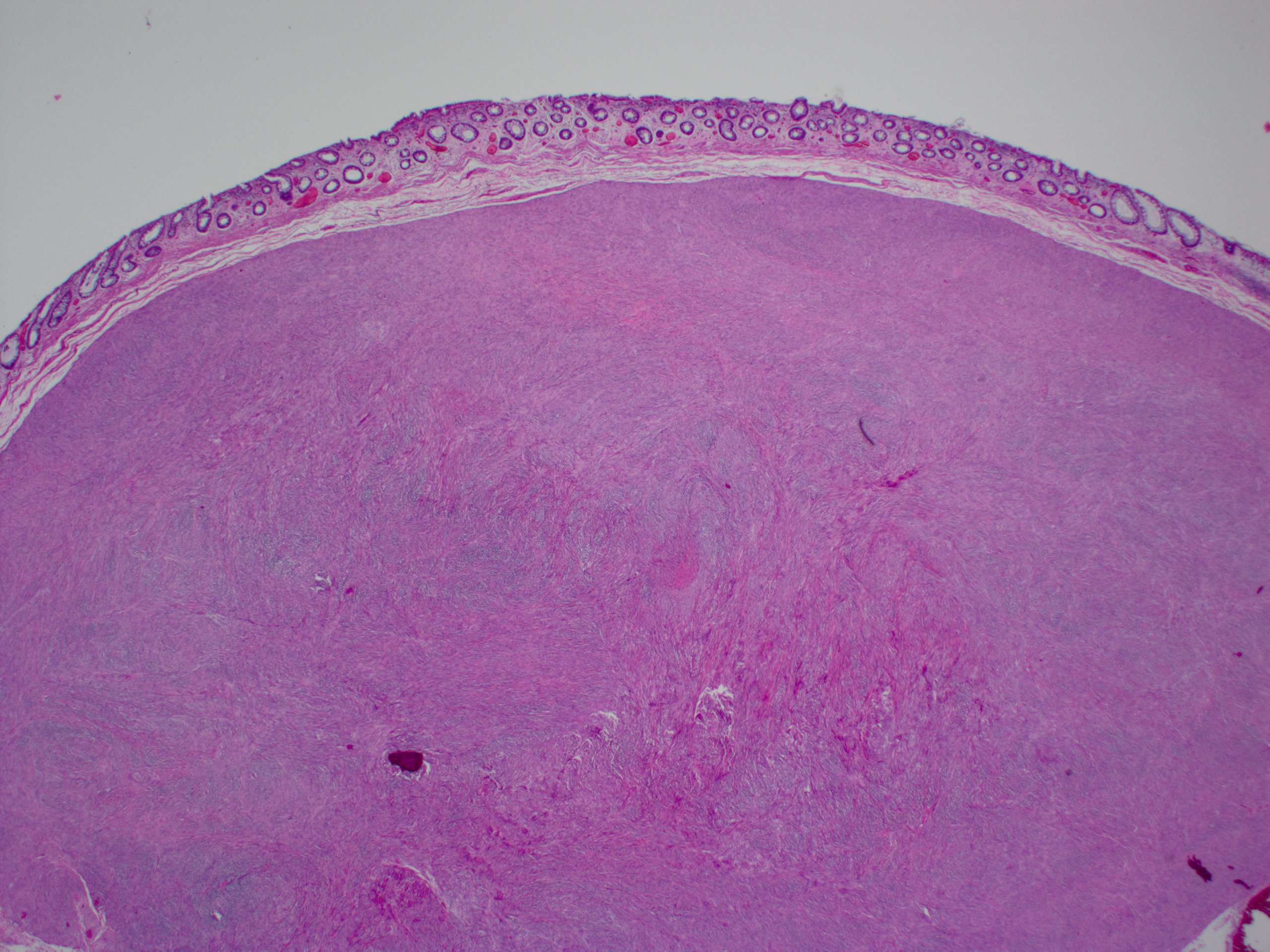
Pathology Outlines - Gastrointestinal stromal tumor

Sex Cord-Stromal and Miscellaneous Tumors of the Ovary - ScienceDirect

Complex Genetic Alterations in Gastrointestinal Stromal Tumors with  Autonomic Nerve Differentiation | Modern Pathology
Complex Genetic Alterations in Gastrointestinal Stromal Tumors with Autonomic Nerve Differentiation | Modern Pathology

Pathology Outlines - Gastrointestinal stromal tumor

A gastric GANT from a 26 year old female showing numerous dense core... |  Download Scientific Diagram
A gastric GANT from a 26 year old female showing numerous dense core... | Download Scientific Diagram

Brendan Dickson, MD on Twitter: "DERMATOFIBROSARCOMA PROTUBERANS. IHC:  +CD34. NB: myxoid area w/ prominent vessels; elsewhere conventional  pattern. https://t.co/8dpMOoB4Kb" / Twitter
Brendan Dickson, MD on Twitter: "DERMATOFIBROSARCOMA PROTUBERANS. IHC: +CD34. NB: myxoid area w/ prominent vessels; elsewhere conventional pattern. https://t.co/8dpMOoB4Kb" / Twitter

jnsclCASE2059 1..5
jnsclCASE2059 1..5

Figure 2 from Histological and immunohistochemical studies on primary  intracranial canine histiocytic sarcomas | Semantic Scholar
Figure 2 from Histological and immunohistochemical studies on primary intracranial canine histiocytic sarcomas | Semantic Scholar

Webpathology.com: A Collection of Surgical Pathology Images

Pathology Outlines - Gastrointestinal stromal tumor

GANT located in distal to the gastroesophageal junction as seen in the... |  Download High-Quality Scientific Diagram
GANT located in distal to the gastroesophageal junction as seen in the... | Download High-Quality Scientific Diagram

ElisePath 🧚🏾‍♀️( Elise Sauvain) (@ElisePath) / Twitter

Figure 5 from Mastoiditis and facial paralysis as initial manifestations of  temporal bone systemic diseases - the significance of the histopathological  examination. | Semantic Scholar
Figure 5 from Mastoiditis and facial paralysis as initial manifestations of temporal bone systemic diseases - the significance of the histopathological examination. | Semantic Scholar

Pleomorphic Sarcomas - Surgical Pathology Clinics

Pathology Outlines - Composite pheochromocytoma